CBCT imaging of the ears
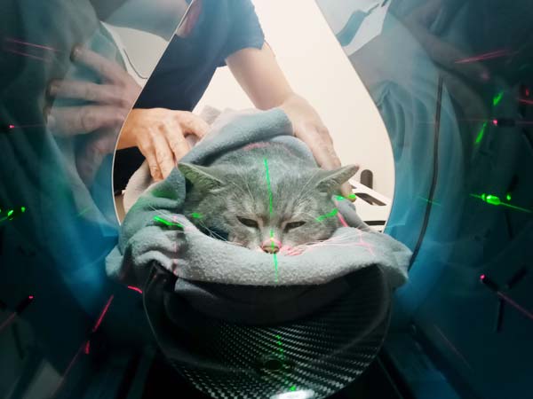
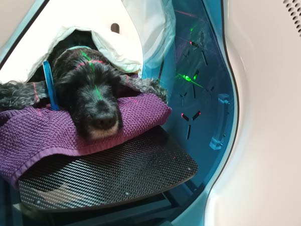
In prolonged ear diseases, an important part of the diagnostics is CBCT imaging of the ears. At Aures we have our own cone beam computed tomography equipment and with this we can get important inormation on how to plan the ear treatment. We usually do the imaging in adjunction to an ear flush and video otoscopy but if needed we can do the imaging separately.
The pros of CBCT imaging
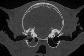
With CBCT imaging we get more information about the ear structures that we can not see with an otoscope or a video otoscope. The most common reason for CBCT imaging is diagnosing or outruling otitis media (middle ear inflammation).
The middle ear is located on the other side of the ear drum and is an ovaloid cavity whose bony walls are covered by a mucous membrane. In the middle ear there are three small bones called the auditory ossicles and they makes the sounds from the ear canal stronger so that the inner ear can perceive them, otherwise a healthy middle ear cavity (bulla) contains only air. From the middle ear there is a channel to the nasopharynx via the Eustachian tube. If there is a problem the middle ear can be completetely or partly filled with inflammatory secretions, slime or soft tissue. Sometimes it is very obvious based on the information given previously to the visit or after the examination at the clinic that the pet has a middle ear problem, but in order to rule out middle ear inflammation or giving a proper assessment CBCT imaging is usually needed.

CBCT imaging of the middle ears is extra important when a cat has had chronic ear problems.
Cone beam computed tomography, CBCT
CBCT imaging is a quick and cost-effective way to outrule middle ear problems and for seeing if the middle ears are filled with pus, fluid or soft tissue or has changes to the bone surrounding them. Magnetic resonance imaging (MRI) and computed tomography (CT) can also be used for imaging ear structures. MRI doesn’t show changes to the bone very well, but it is superior for assessment of soft tissue changes. This is why MRI is recommended for assessment of tumours or when a pet has severe neurologic symtoms. In CT images the bony structures are easily seen and soft tissue is distinguished more easily than with CBCT (especially when a contrast agent is used), but its high cost is often a problem, particularly for ruling out middle ear disease.

In addition to otoscopy, imaging is helpful for choosing the right treatment for ear inflammations in cats and dogs.
Middle ear inflammation (otitis media)
Middle ear inflammation is mostly associated with chronic or severe outer ear inflammations. In these cases the ear drum has been damaged and the inflammation has spread to the middle ear. Holes in the eardrum can heal pretty quickly, and because of this an intact ear drum doesn’t rule out middle ear inflammation. Dogs can also have middle ears problem that are caused by their anatomy and are not necessarily linked to an inflammation, this can be seen in dog breeds with very round skulls.
Middle ear inflammations in dogs are usually treated with orally administrated drugs, video otoscopy assisted flushing of the middle ear and topical treatment of the ear.
Inflammatory changes to the bony wall surrounding the middle ear usually need to be treated with surgery. This is one of the most important reasons to we do CBCT imaging of all our ear patients that are here for a video otoscopy – if the problem is not treatable without surgery, it is important to know about this at an early stage, so that the painful ear problem is not prolonged and the owner’s money isn’t used for treatments that will not provide a permanent cure.
Flushing the middle ear of cats is challenging because of the bony wall in their middle ear. The wall must be punctured in order to examine and treat the whole middle ear.
Because cats are very good at hiding their pain, the response to treatment can not be reliably assessed based on the cat’s behaviour. The response to treatment is therefore followed up with CBCT imaging.

Prolonged ear inflammations in dogs may often lead to middle ear inflammation. Imaging of the middle ears is an important part of the diagnostics.
